
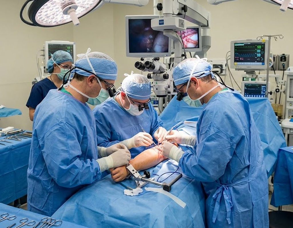
Sarcomas and other advanced tumours may surround, invade or adhere to major limb vessels. In selected patients, en bloc tumour resection with planned vascular reconstruction can achieve an appropriate oncologic operation while preserving the limb. These cases require detailed imaging and close coordination between oncologic, vascular and reconstructive teams.
Contrast-enhanced MRI defines the tumour and soft tissues, while CT angiography, MR angiography or duplex imaging may assess arterial and venous anatomy. Planning considers collateral circulation, proximal and distal landing zones, potential conduit, soft-tissue coverage, anticipated ischaemia time and the need for nerve or bone reconstruction.
A resected artery may be reconstructed using the patient’s own vein or a prosthetic vascular graft, depending on vessel size, required length, available conduit, contamination and surgeon judgement. Venous reconstruction is considered selectively. Healthy vascularised soft-tissue coverage may protect the graft and fill dead space.
Antiplatelet or anticoagulant treatment is individualised according to the reconstructed vessel, conduit, bleeding risk, flow and treating team’s protocol. No website should promise one lifelong regimen for every graft. Clinical examination and duplex or cross-sectional imaging may be used for surveillance.
Potential complications include early or late thrombosis, bleeding, infection, graft exposure, embolism, limb ischaemia, oedema, wound failure, tumour recurrence and need for revision or amputation. Risk is influenced by tumour extent, contamination, conduit, runoff, radiotherapy and patient factors.
copyright © 2026, All rights reserved | Dr Saurabh Gupta